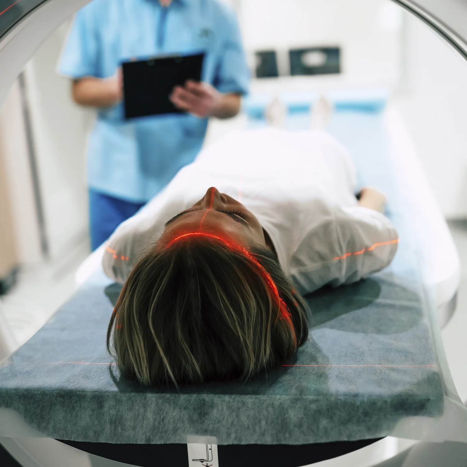
Magnetic Resonance Imaging (MRI) is a medical imaging technique that uses powerful magnets, radio waves, and a computer to generate detailed images of the internal structures of the body. The development of MRI is a fascinating story that involves contributions from multiple researchers over several decades. Here is a brief history of MRI:
- Discovery of Nuclear Magnetic Resonance (NMR):
- The foundation for MRI was laid in the 1940s with the discovery of Nuclear Magnetic Resonance (NMR). In 1946, Felix Bloch and Edward Mills Purcell independently discovered the phenomenon of NMR, for which they were awarded the Nobel Prize in Physics in 1952. NMR is based on the behavior of atomic nuclei in a magnetic field.
- Development of NMR for Medical Imaging:
- In the 1970s, scientists began exploring the use of NMR for medical imaging. Paul Lauterbur, an American chemist, introduced the concept of using magnetic field gradients to create spatial images. In 1973, he published a paper describing the principles of MRI and demonstrated the first two-dimensional image using NMR. Lauterbur’s contributions earned him a share of the Nobel Prize in Physiology or Medicine in 2003.
- Independent Contributions:
- Around the same time, Sir Peter Mansfield, a British physicist, was independently working on improving MRI techniques. In the late 1970s and early 1980s, Mansfield made significant advancements in the development of rapid imaging techniques, such as echo-planar imaging (EPI), which allowed faster image acquisition.
- Clinical Applications:
- In the 1980s, MRI technology began to be applied to clinical medicine. The ability of MRI to provide detailed images of soft tissues without the use of ionizing radiation made it a valuable tool for diagnosing a wide range of medical conditions.
- Advancements and Innovations:
- Over the years, there have been numerous advancements and innovations in MRI technology, including improvements in image resolution, faster scan times, and the development of specialized MRI techniques for different types of tissues and diseases.
- Functional MRI (fMRI):
- In the 1990s, functional MRI (fMRI) emerged as a powerful tool for mapping brain activity by measuring changes in blood flow. This opened up new possibilities for studying the brain’s function and connectivity.
- Ongoing Developments:
- MRI technology continues to evolve, with ongoing research aimed at improving image quality, reducing scan times, and exploring new applications. Recent developments include the use of artificial intelligence to enhance image reconstruction and interpretation.
Today, MRI is a widely used imaging modality in medical practice, providing valuable diagnostic information across various medical specialties. Its non-invasive nature and ability to visualize soft tissues in detail make it an indispensable tool in modern healthcare.


