


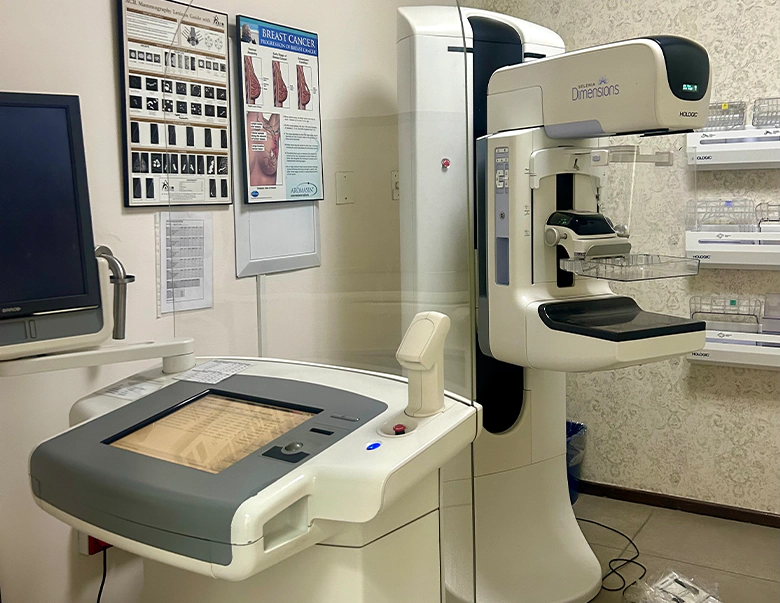
Mammography is a special type of imaging that uses a low-dose X-ray system to examine breasts. A mammography exam, called a mammogram is used to aid in the early detection and diagnosis of breast disease in women.
Digital breast tomosynthesis mammography, also known as 3D mammography, is an advanced form of mammography that captures multiple images of the breast from different angles. These images are then reconstructed into a detailed 3D view of the breast tissue. The primary goal of tomosynthesis is to provide a clearer and more accurate assessment of breast abnormalities, especially for women with dense breasts. It helps in detecting smaller tumours and reducing false-positive results that can occur with traditional 2D mammography. Combining digital breast tomosynthesis mammography and breast ultrasound can provide a comprehensive evaluation of breast tissue.
Is booking required?
Yes
Branches: Mediclinic Vereeniging and Midvaal Private Hospitals
Facts about breast cancer
- Although most women are diagnosed between the ages of 45 and 70 years, it can also occur in younger women less than 45 years of age.
- The earlier breast cancer is diagnosed, the better the prognosis of survival and quality of life.
- There is a 90% 5 year survival rate when breast cancer is detected before it spreads.
- All breast lumps should be investigated; 9/10 lumps are not malignant.
Factors that can put you at an increased risk for breast cancer
- Age: More likely women over 50 years, but can also be found in a young woman.
- Gender: Women are more likely to be affected than men.
- Family history: Having close relatives with breast cancer.
- If you had breast cancer in one breast, you have an increased risk of getting breast cancer in the other breast.
- Menstrual history: Early onset of menstruation (before 12 years) and late menopause (after 50 years)
- Children: Having no children or having your first child after the age of 30 years
- HRT: Taking hormone replacement therapy for more than 5 years.
- Lifestyle: Smoking, alcohol, obesity and a high fat diet are all harmful and can increase the risk of cancer.
Value of screening mammography
- Early detection: Mammogram can detect a mass up to 2 years before detection by palpation.
- Enables more successful treatment options: Lumpectomy (removal of a lump) instead of mastectomy (removal of breast), so early detection can save your breast and more importantly, your life.
- Trials have shown mortality reduction of up to 40-45% in ages 50-70 years.
- Screening mammography: X-ray of the breast with no symptoms.
- Diagnostic mammogram: X-ray of the breast where a known problem exists or where the patient has symptoms.
How should I prepare for the procedure?
- Do not apply powder, deodorants and creams to your chest area.
- Try not to schedule your mammogram for the week before or during your menstrual period as your breasts are generally more sensitive during this time.
- Please inform the radiographer if you are pregnant or suspect that you may be pregnant.
- Any breast symptoms or problems must be discussed with the radiographer performing the examination.
What does the procedure entail?
The breast is compressed between 2 plates and specialised x-ray machine moves in an arc over the breast taking multiple images in a matter of seconds. The process may cause some temporary discomfort due to the pressure applied but is generally well tolerated. The obtained images are then reviewed by a radiologist who can examine each layer of the breast tissue individually, enhancing their ability to detect and characterise abnormalities. Ultrasound is used as a complementary imaging modality to mammography, especially for further evaluation of suspicious findings or dense breasts. Breast ultrasound can help differentiate between solid masses and fluid filled cysts, guide biopsy procedures and assess blood flow in breast lesions. Stereotactic and ultrasound guided vacuum assisted breast biopsies are a type of biopsy under local anaesthesia which uses special instrument which utilises vacuum suction and therefore enables more tissue to be removed. The examination usually takes approximately 20-40 minutes. Your results will be sent to the referring doctor. Please contact your doctor to discuss the results if they have not contacted you in a timely manner.

Thank you for considering Vaal Radiology for your diagnostic imaging needs. We look forward to serving you soon.


Available Doctors
Dr MPG Kitsa
Dr AB Mehtar
Dr VJ Radebe
Dr J Veldman
Midvaal Private Hospital
Tel: 016 430 4330
Nile Drive Three Rivers
Accounts
Tel: 016 430 4350/4357
Email: accounts@vaalrad.co.za
PO Box 838 Vereeniging 1930
Mediclinic Vereeniging
Tel: 016 430 4300
Cnr Joubert/Rhodes Ave Vereeniging

Dr MPG Kitsa
Dr AB Mehtar
Dr VJ Radebe
Dr J Veldman
Tel: 016 430 4330
Nile Drive Three Rivers
Tel: 016 430 4350/4357
Email: accounts@vaalrad.co.za
PO Box 838 Vereeniging 1930
Tel: 016 430 4300
Cnr Joubert/Rhodes Ave Vereeniging





Proudly designed by Bunnypants Graphic & Web Design Studio | 2024